
Based on PubMed | In head and neck cancer, what can cause tingling in the face, tongue, or throat, and how does this symptom relate to prognosis?
Tingling or numbness in the face, tongue, or throat in head and neck cancer can stem from perineural invasion/spread, cranial nerve or skull base involvement, mass effect, inflammation/infection, or treatment-related neuropathy (radiation or chemotherapy). When caused by perineural invasion or cranial neuropathy, it often indicates more advanced disease and worse prognosis with higher recurrence risk; treatment-related tingling reflects toxicity rather than progression. Persistent focal paresthesias warrant a cranial nerve exam and contrast-enhanced MRI to assess perineural pathways.
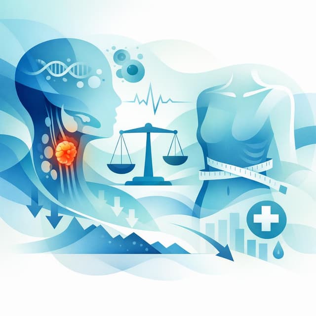
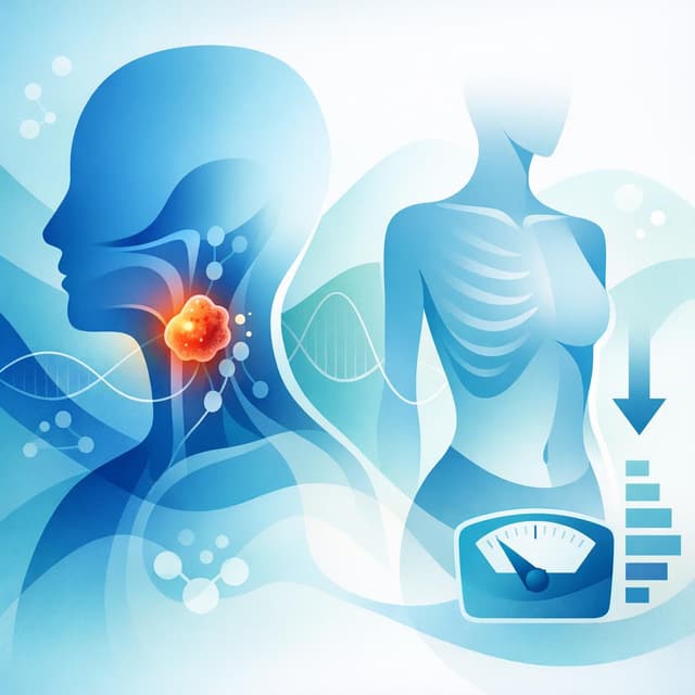
Head and neck cancer can cause tingling, numbness, or “pins and needles” in the face, tongue, or throat for several reasons, and in some situations this symptom can signal more-aggressive disease or a higher risk of recurrence. Most commonly, tingling arises from tumor-related nerve involvement (perineural invasion or spread), treatment-related nerve injury (radiation or chemotherapy), inflammation or infection, or mechanical compression from tumors or lymph nodes. [1] [2] [3] [4] [5] [6] [7] [8] [9]
Key causes of tingling
-
Perineural invasion/spread (PNI/PNS): Cancer cells can grow along nerves, especially the trigeminal (cranial nerve V) and facial (cranial nerve VII), causing facial or oral tingling, numbness, or pain. PNI is a recognized pathway of tumor extension in head and neck cancers and is best evaluated with MRI, which can show nerve enlargement and abnormal enhancement. [1] Trigeminal neuropathy from perineural spread often presents with facial pain or paresthesias (tingling) and can be confirmed with contrast-enhanced MRI. [10] PNI occurs across head and neck squamous cell carcinomas and salivary and cutaneous primaries and is associated with worse local control and survival in many studies. [3] [11]
-
Direct cranial nerve involvement or skull base invasion: Tumors of the mouth, oropharynx, sinonasal region, or salivary glands can infiltrate or compress cranial nerves, leading to sensory changes such as tingling or numbness in the lips, cheeks, tongue, or palate. Symptoms like mouth numbness and trouble moving the tongue are recognized signs in certain oral cavity cancers. [12] Imaging with MRI and CT helps define soft tissue and bony/skull base involvement. [13] [14]
-
Radiation-induced neuropathy: Radiation to the head and neck can inflame or damage nerves over time, leading to sensory changes including tingling, often months to years after treatment. Head and neck radiation commonly causes mucosal symptoms (mouth and throat soreness, swallowing pain, dry mouth), and neuropathic symptoms may occur as a late effect that warrants clinician evaluation. [15] [16] [4] [5]
-
Chemotherapy-related peripheral neuropathy: Drugs used in some head and neck regimens (for example, platinum agents like cisplatin and certain combinations) can cause tingling or “pins and needles,” usually starting in the hands and feet but sometimes felt around the mouth. Patients are advised to report new tingling, numbness, or pain in extremities or around the mouth during therapy, as this may indicate treatment-related nerve effects. [7] These agents are known to cause peripheral neuropathy that may interfere with daily tasks. [6]
-
Inflammation, infection, and post-treatment scarring: Severe mucositis, post-surgical changes, fibrosis, or infections can irritate sensory nerves and produce transient tingling or numbness, particularly in the mouth and throat. Persistent sores, ulcers, or masses that fail to heal in the mouth and throat remain red-flag symptoms and should be assessed. [17] [9] [8]
-
Nodal or mass effect (compression): Enlarged lymph nodes or tumors in confined spaces (e.g., pterygopalatine fossa, skull base foramina) can press on sensory branches and cause localized tingling. Neck lumps and masses are common in head and neck cancers and may accompany other sensory changes. [17] [9] [8]
Why tingling matters for prognosis
-
Perineural invasion is a negative prognostic marker: Across head and neck squamous cell carcinoma, PNI is linked to significantly worse outcomes, including overall survival, disease-free survival, and higher risks of local and locoregional recurrence and distant spread. A large meta-analysis showed PNI is associated with substantially poorer overall survival and increased local and regional recurrence. [3] This makes tingling from PNI particularly important to recognize and stage accurately. [3]
-
Clinical (symptomatic) perineural disease tends to do worse than microscopic PNI: When nerve involvement causes symptoms like numbness or tingling (as opposed to being found only under the microscope), outcomes for local control and disease-free survival tend to be poorer, underscoring the need for aggressive evaluation and tailored therapy. Comparative data in cutaneous head and neck carcinomas show lower control rates and higher complication risks when PNI is clinically apparent. [2]
-
Cranial neuropathy from perineural spread indicates advanced local extension: Trigeminal neuropathy with facial paresthesias due to perineural spread often reflects tumor tracking toward the skull base, which can complicate surgical clearance and radiotherapy targeting and may portend more challenging disease control. [10] [1]
-
Treatment-related neuropathy and prognosis: When tingling is due to chemotherapy or radiation rather than tumor, it typically reflects treatment toxicity rather than cancer progression and does not, by itself, worsen cancer prognosis. [6] [7] However, persistent or worsening symptoms still merit evaluation to distinguish toxicity from recurrence. [4] [5]
What to look for clinically
-
Pattern and location of tingling: Facial distribution following trigeminal branches (V2 upper cheek/upper lip; V3 lower jaw/lip; V1 forehead) suggests cranial nerve involvement or PNI, whereas stocking–glove patterns suggest chemotherapy neuropathy. Trigeminal neuropathy due to malignant invasion often aligns with specific nerve divisions and can be visualized on MRI with contrast and fat suppression. [1] [10]
-
Associated signs: New pain, progressive numbness, muscle weakness, difficulty chewing, tongue deviation, or changes in speech/swallowing raise concern for cranial nerve dysfunction or skull base involvement. Oral cavity and oropharyngeal cancers can present with numbness, trouble moving the tongue, sore throat, and swallowing difficulty. [12] Persistent sores or nonhealing lesions in the mouth and throat remain warning signs. [17] [9]
-
Timing relative to therapy: Tingling that begins during or soon after platinum-based chemotherapy may indicate drug-induced neuropathy, while symptoms emerging months to years after radiation may point to radiation neuropathy or recurrence; both require clinical correlation and imaging when persistent. Patients receiving these treatments are advised to report new tingling promptly. [7] [6] [4] [5]
Recommended diagnostic workup
-
Focused head and neck exam and cranial nerve testing: Map sensory changes and screen for motor deficits or mucosal lesions. General symptom sets for head and neck cancer include lumps, nonhealing sores, sore throat, swallowing issues, and voice changes that guide targeted evaluation. [17] [9]
-
High-resolution MRI with contrast of the skull base and perineural pathways: MRI is the preferred modality to detect perineural spread or nerve involvement because it shows direct signs (nerve enlargement, enhancement) and indirect signs (denervation atrophy, loss of fat planes). Fat-suppressed, contrast-enhanced sequences are considered essential for delineating perineural disease. [1] MRI is widely used to detect soft-tissue extension of head and neck tumors and to evaluate abnormal findings. [13] [18]
-
CT for bony foramina and skull base: CT complements MRI by identifying foraminal enlargement, erosion, or other bone changes caused by tumor spread along nerves. CT and MRI together refine staging and radiotherapy fields. [1] [14]
-
PET-CT when recurrence or distant spread is suspected: Whole-body metabolic imaging can assess for regional and distant disease, informing prognosis and treatment planning. [19]
-
Biopsy when feasible: Confirm recurrence or perineural invasion pathologically if imaging suggests tumor along nerve pathways or in new lesions. [2] Pathology helps differentiate treatment effect from viable tumor and guides adjuvant therapy decisions. [2]
Management implications
-
Escalated local therapy for PNI: Because PNI raises recurrence risk, treatment plans often include wider surgical margins when possible and adjuvant radiotherapy that specifically covers involved nerve pathways to the skull base. Outcomes are better for microscopic PNI than for symptomatic/clinical PNI, emphasizing the benefit of early detection and comprehensive targeting. [2] Advanced radiation techniques can improve coverage of complex perineural routes. [11]
-
Systemic therapy for recurrent or advanced disease: If tingling reflects locoregionally advanced or recurrent cancer, systemic therapy may be indicated based on tumor site, stage, and biomarkers, in combination with local treatments. Comprehensive staging with MRI and PET-CT helps select the appropriate approach. [19] [13]
-
Supportive care for treatment-induced neuropathy: For chemotherapy-induced neuropathy, treatment adjustments, symptom control strategies, and rehabilitation may be considered; for radiation-related neuropathy, symptom management and surveillance imaging help differentiate late effects from recurrence. Patients are encouraged to promptly report new tingling during therapy to enable early mitigation. [7] [6] [4] [5]
Quick reference: causes and prognostic meaning
| Cause | Typical features | How it relates to prognosis |
|---|---|---|
| Perineural invasion/spread | Facial or oral tingling/numbness in trigeminal distribution; MRI shows nerve enhancement/enlargement | Associated with worse survival and higher local/locoregional recurrence; symptomatic (clinical) PNI has poorer control than microscopic PNI. [1] [10] [3] [2] |
| Direct cranial nerve or skull base involvement | Focal sensory loss, pain, or motor deficits; CT/MRI define extent | Often indicates more-advanced local disease, requiring comprehensive local therapy and careful staging. [13] [14] |
| Chemotherapy-induced neuropathy (e.g., platinum) | Stocking–glove tingling; may include peri-oral sensations; emerges during/after treatment | Toxicity marker rather than progression; prompts dose review and symptom management. [7] [6] |
| Radiation-induced neuropathy | Delayed onset after radiotherapy; focal or diffuse sensory changes | Late treatment effect; not inherently a worse cancer prognosis but must be distinguished from recurrence. [4] [5] |
| Mass effect from tumor or nodes | Tingling near a palpable mass; imaging shows compression | Suggests active disease burden; prognosis depends on stage and response to therapy. [17] [9] |
Take-home message
Tingling in the face, tongue, or throat during the course of head and neck cancer care has several potential causes, but new or progressive numbness/tingling localized to the face or oral cavity can be a red flag for perineural invasion or cranial nerve involvement, which is generally linked to a higher risk of recurrence and poorer survival compared to cases without PNI. [3] [1] Because MRI is the best test to detect perineural spread, persistent or focal paresthesias should usually prompt a targeted cranial nerve exam and contrast-enhanced MRI of the skull base and perineural pathways, with CT and PET-CT as needed. [1] [13] [19] On the other hand, tingling that starts with chemotherapy or appears long after radiation may reflect treatment effects rather than tumor progression, but it still deserves evaluation to be safe. [7] [6] [4] [5]
Related Questions
Related Articles

Sources
- 1.^abcdefghiTrigeminal perineural spread of head and neck tumors.(pubmed.ncbi.nlm.nih.gov)
- 2.^abcdefCutaneous head and neck basal and squamous cell carcinomas with perineural invasion.(pubmed.ncbi.nlm.nih.gov)
- 3.^abcdefThe Prognostic Role of Perineural Invasion for Survival in Head and Neck Squamous Cell Carcinoma: A Systematic Review and Meta-Analysis.(pubmed.ncbi.nlm.nih.gov)
- 4.^abcdefgRadiation Therapy to Your Head and Neck(mskcc.org)
- 5.^abcdefgRadiation Therapy for Head and Neck Cancer(mskcc.org)
- 6.^abcdefgPatient information - Head and neck cancer recurrent or metastatic - Cisplatin, fluorouracil and pembrolizumab(eviq.org.au)
- 7.^abcdefgPatient information - Head and neck cancer recurrent or metastatic - Cisplatin, fluorouracil and pembrolizumab(eviq.org.au)
- 8.^abcHead and Neck Cancer Symptoms & Signs | Memorial Sloan Kettering Cancer Center(mskcc.org)
- 9.^abcdefHead and Neck Cancer(medlineplus.gov)
- 10.^abcdTrigeminal neuropathy secondary to perineural invasion of head and neck carcinomas.(pubmed.ncbi.nlm.nih.gov)
- 11.^abPerineural invasion and spread in head and neck cancer.(pubmed.ncbi.nlm.nih.gov)
- 12.^abTypes of Head & Neck Cancer(nyulangone.org)
- 13.^abcdeDiagnosing Head & Neck Cancer(nyulangone.org)
- 14.^abcDiagnosing Head & Neck Cancer(nyulangone.org)
- 15.^↑Radiation Therapy to Your Head and Neck(mskcc.org)
- 16.^↑Radiation Therapy Side Effects(mskcc.org)
- 17.^abcdeHead and neck cancers - Symptoms and causes(mayoclinic.org)
- 18.^↑Imaging Tests for Head and Neck Cancer(stanfordhealthcare.org)
- 19.^abcDiagnosing Head & Neck Cancer(nyulangone.org)
Important Notice: This information is provided for educational purposes only and is not intended to replace professional medical advice, diagnosis, or treatment. Always consult with a qualified healthcare provider before making any medical decisions.