Based on PubMed | What do elevated liver enzymes indicate in gallbladder cancer, and which patterns of AST, ALT, ALP, and GGT elevation help differentiate biliary obstruction from liver metastases?
Elevated liver enzymes in gallbladder cancer usually reflect either cholestasis from biliary obstruction or liver involvement from invasion or metastases. A cholestatic pattern with disproportionately high ALP/GGT and bilirubin suggests obstruction, while metastases more often cause ALP/GGT elevations with only mild AST/ALT increases; isolated aminotransferase spikes point to hepatocellular injury. Enzymes are supportive, not diagnostic, so imaging is required.
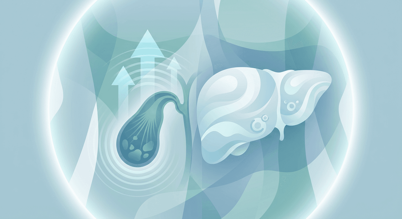
Elevated liver enzymes in gallbladder cancer often signal either blockage of bile flow (cholestasis) from tumor-related obstruction or injury/infiltration of the liver from direct invasion or metastases. Elevations can also reflect coexisting conditions like hepatitis or fatty liver, so the pattern across multiple tests AST, ALT, alkaline phosphatase (ALP), gamma‑glutamyl transferase (GGT), and bilirubin helps narrow the cause. High bilirubin and cholestatic enzymes (ALP, GGT) commonly rise with biliary obstruction, while liver metastases more often raise ALP and GGT and may raise AST/ALT modestly; isolated aminotransferase spikes (AST/ALT) point more to hepatocellular injury than obstruction. [1] [2] Elevated bilirubin suggests impaired bile flow or liver function, which can occur in gallbladder cancer; liver enzyme tests (ALP, AST, ALT, GGT) and tumor markers (CEA, CA 19‑9) are part of work‑ups but are not diagnostic on their own. [3]
Why enzymes rise in gallbladder cancer
- Biliary obstruction: A tumor in the gallbladder, cystic duct, common bile duct, or hilar region can obstruct bile, causing cholestasis; this typically elevates ALP, GGT, and bilirubin more than AST/ALT. [1] [2]
- Liver involvement/metastases: Tumor spread to the liver parenchyma can injure bile canaliculi and hepatocytes, commonly elevating GGT and ALP, with mild–moderate AST/ALT increases; normal enzymes do not exclude metastases. [4] [5]
Typical lab patterns
- Cholestatic pattern (obstruction): Disproportionately high ALP and GGT compared with AST/ALT, often with elevated bilirubin. [1] [2]
- Hepatocellular pattern: Disproportionately high AST and ALT relative to ALP; this is less typical of pure extrahepatic obstruction and suggests primary hepatocyte injury. [6] [7]
- Mixed pattern: Both aminotransferases and cholestatic enzymes are elevated; can be seen with liver metastases or intrahepatic cholestasis from tumor infiltration. [4] [5]
Enzyme-by-enzyme clues
- ALP: Elevates with bile duct obstruction and with space‑occupying liver lesions, including metastases; useful for detecting lesions that cause cholestasis. [1]
- GGT: Sensitive to cholestasis and liver metastases; elevations are common in metastatic liver disease and in jaundice; helps confirm hepatic source of ALP. [4] [8] [2]
- AST/ALT: Often modestly elevated in liver metastases; isolated marked aminotransferase surges favor hepatocellular injury over pure obstruction. [4] [6] [7]
- Bilirubin: Elevates in obstruction or significant hepatic dysfunction; high levels with ALP/GGT suggest cholestasis from obstructive gallbladder or biliary lesions. [3] [1]
Evidence summaries
- In sonographically confirmed liver metastases, GGT and ALP were elevated in ~89% and ~88% of cases, respectively; AST and ALT rose less often, and normal enzymes did not rule out metastases. [4]
- In gastrointestinal cancers without obvious hepatic involvement, ALP and GGT were the most sensitive and specific biochemical tests for hepatic metastases; abnormal combinations (e.g., GGT with LDH or AST) improved predictive value. [5]
- GGT tends to be elevated in nearly all jaundiced states and is frequently elevated in liver metastases even without jaundice, making it a helpful marker when ALP is high. [8]
Practical differentiation: obstruction vs metastasis
| Pattern feature | Biliary obstruction (cholestasis) | Liver metastases |
|---|---|---|
| ALP | Markedly elevated (disproportionate) | Commonly elevated |
| GGT | Markedly elevated | Commonly elevated, often sensitive |
| Bilirubin | Often elevated (especially if extrahepatic blockage) | May be normal or elevated, depends on extent/duct involvement |
| AST/ALT | Mild–moderate rise, less than ALP/GGT | Mild–moderate rise; AST/ALT often lower relative to GGT/ALP |
| Overall pattern | Cholestatic | Mixed cholestatic with possible mild hepatocellular component |
| Caveat | Does not alone prove obstruction; needs imaging | Normal enzymes do not exclude metastases |
Sources for table elements: ALP indicates lesions causing biliary obstruction; 5′‑nucleotidase and GGT support cholestasis; bilirubin rises with impaired bile flow. [1] [2] GGT and ALP are highly sensitive in liver metastases; AST/ALT elevations are less frequent, and normal enzymes may still occur in metastases. [4] [5] GGT rises with jaundice and is suggestive of metastases when elevated without jaundice. [8]
How clinicians put it together
- Combine enzyme patterns with symptoms (e.g., jaundice, dark urine, pale stools, itching), physical exam, and imaging (ultrasound, CT/MRI, MRCP/ERCP). Imaging is usually required to confirm obstruction or detect metastases. [3]
- Use cholestatic markers to guide urgency: markedly high ALP/GGT with rising bilirubin and cholangitic symptoms often prompt expedited biliary imaging and possible decompression. [1] [2]
- Recognize limitations: Enzymes are supportive, not definitive; overlap is common, and mixed patterns occur with intrahepatic cholestasis from metastatic infiltration. [4] [5]
Additional notes
- Ancillary markers like 5′‑nucleotidase can help confirm hepatic origin of ALP when bone disease is a concern. [2]
- Leucine aminopeptidase (LAP) has been reported to rise broadly in hepatobiliary malignancies, sometimes outperforming other enzymes, though it is less commonly used in modern practice. [9]
Key takeaways
- Predominant ALP/GGT ± bilirubin elevation suggests cholestasis from biliary obstruction; disproportionate AST/ALT suggests primary hepatocellular injury; metastases often produce a mixed cholestatic pattern with ALP/GGT most sensitive. [1] [2] [4] [5] [8]
- Normal enzymes do not exclude liver metastases, so imaging remains essential when suspicion is high. [4]
Would you like to share your most recent liver test values (AST, ALT, ALP, GGT, bilirubin) so I can help interpret the pattern in your specific situation?
Related Questions
Related Articles


Sources
- 1.^abcdefghLiver function tests(stanfordhealthcare.org)
- 2.^abcdefghLiver function tests(stanfordhealthcare.org)
- 3.^abcGallbladder Cancer Diagnosis(mskcc.org)
- 4.^abcdefghi[Pathologic enzyme patterns in sonographically verified liver metastases].(pubmed.ncbi.nlm.nih.gov)
- 5.^abcdefHepatic metastases in gastrointestinal cancer: diagnostic value of biochemical investigations.(pubmed.ncbi.nlm.nih.gov)
- 6.^abLiver function tests(stanfordhealthcare.org)
- 7.^abElevated liver enzymes - Mayo Clinic(mayoclinic.org)
- 8.^abcdGamma-glutamyl transpeptidase as an aid in the diagnosis of liver metastases.(pubmed.ncbi.nlm.nih.gov)
- 9.^↑Leucine amino peptidase a better indicator of carcinoma of liver, biliary tract and pancreas.(pubmed.ncbi.nlm.nih.gov)
Important Notice: This information is provided for educational purposes only and is not intended to replace professional medical advice, diagnosis, or treatment. Always consult with a qualified healthcare provider before making any medical decisions.