Based on PubMed | What symptoms and evaluation steps belong on a patient checklist for unintentional weight loss related to bile duct cancer?
This patient checklist links unintentional weight loss to potential bile duct cancer by tracking key symptoms (jaundice, dark urine, pale stools, itching, RUQ pain, anorexia/fatigue) and risk factors (PSC, cirrhosis, biliary disease). It provides a stepwise evaluation history/exam, labs (LFTs, CBC, CA 19-9/CEA), imaging (ultrasound to CT/MRI/MRCP), and procedures (EUS-FNA, ERCP/PTC) plus nutrition support and urgent-care red flags.
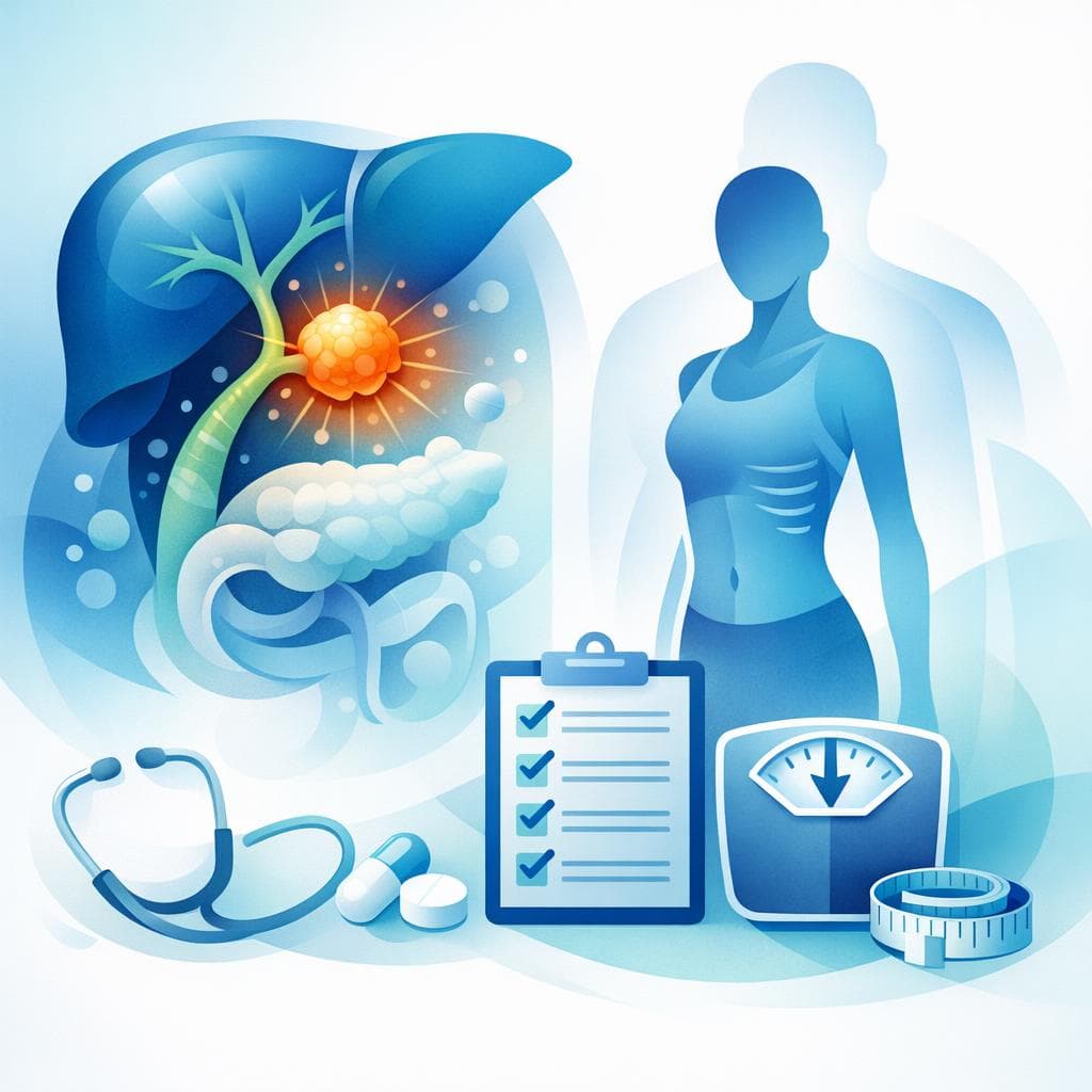
Unintentional Weight Loss and Bile Duct Cancer: What to Include in a Patient Checklist
Unintentional weight loss can be a warning sign of many conditions, including bile duct cancer (cholangiocarcinoma). To help users and clinicians recognize important symptoms early and organize next steps, the following checklist brings together evidence-based symptoms, risk factors, and a stepwise evaluation plan, including history, examination, labs, imaging, and when to consider tissue diagnosis. Because early symptoms are often subtle, a structured checklist can support timely evaluation. [1] [2]
Key Symptoms to Track
-
Jaundice (yellowing of skin/eyes) and dark urine
-
Pale, clay-colored, or greasy stools
- Light or clay-colored stools suggest reduced bile reaching the intestine. [3]
-
Itchy skin (pruritus)
-
Right-upper-quadrant or upper abdominal pain, possibly radiating to the back
-
Nausea, vomiting, and loss of appetite
-
Unintentional weight loss and fatigue
-
Fever or chills (if infection or cholangitis is present)
- May suggest superimposed infection on biliary obstruction and needs urgent care. [5]
Tip: Record the onset date, severity, and daily pattern of each symptom. Tracking symptom progression (especially jaundice and stool/urine color changes) helps prioritize urgent testing. [1] [3]
Who Is at Higher Risk?
-
Primary sclerosing cholangitis (PSC), chronic biliary inflammation, and certain liver diseases (e.g., cirrhosis)
-
History of bile duct strictures, gallstones, pancreatitis, or prior biliary surgery
- These may cause scarring/strictures associated with symptoms similar to malignancy. [5]
-
Inflammatory bowel disease (particularly ulcerative colitis) through association with PSC
- PSC often coexists with ulcerative colitis and increases cholangiocarcinoma risk. [1]
Action: Include known liver/bile duct conditions and prior biliary procedures in the checklist’s risk section. [1] [5]
Stepwise Evaluation: From History to Diagnosis
A structured approach improves diagnostic yield and safety. This stepwise pathway prioritizes non-invasive tests first and uses targeted invasive methods for confirmation and symptom relief. [1] [2] [6]
1) History
- Weight loss details: total amount, timeframe, pace, and diet changes
- Unintentional weight loss is defined as ~5% of body weight over 6–12 months. [7]
- Symptom inventory: jaundice, dark urine, pale stools, itching, RUQ pain, nausea/vomiting, fever/chills
- Past medical history: PSC, cirrhosis, gallstones, pancreatitis, prior ERCP or surgery
- Medication and social history: alcohol use, hepatotoxic drugs, and overall nutritional intake
2) Physical Examination
- General: weight trend, signs of malnutrition
- Skin/eyes: jaundice and scratch marks (from pruritus)
- Abdomen: right-upper-quadrant tenderness, hepatomegaly
- Hepatomegaly can accompany biliary obstruction or malignancy. [10]
3) Initial Laboratory Tests
- Liver function tests: bilirubin, alkaline phosphatase (ALP), AST/ALT
- Cholestasis often shows elevated bilirubin and ALP. [11]
- Complete blood count (CBC)
- Screens for anemia, infection. [11]
- Basic metabolic panel and inflammatory markers as needed
- Broader weight loss workup includes systemic checks. [12]
- Tumor markers: CA 19-9 and CEA (supportive, not definitive)
4) First-Line Imaging
- Abdominal ultrasound
- Useful to detect bile duct dilation, gallbladder pathology, or mass effect; often first imaging step. [2]
- Contrast-enhanced CT or MRI/MRCP for characterization and staging
5) Targeted Diagnostic Procedures (if obstruction/mass suspected)
- Endoscopic ultrasound (EUS) with fine-needle aspiration (FNA)
- Helpful for tissue sampling near the bile duct and pancreatic head. [2]
- Endoscopic retrograde cholangiopancreatography (ERCP) with brushings/biopsy and possible stent placement
- Allows tissue sampling and therapeutic biliary drainage for symptomatic obstruction. [2]
- Percutaneous transhepatic cholangiography (PTC) when ERCP is not feasible
- Provides biliary mapping and access for drainage in selected cases. [10]
Note: Tissue diagnosis confirms malignancy, but sensitivity of brushings/biopsies can be limited; multiple techniques may be needed. [6]
Differential Diagnosis to Consider
- Benign bile duct stricture or gallstone-related obstruction
- Can produce similar symptoms: jaundice, itching, pale stools, RUQ pain, fever. [5]
- Pancreatic or small intestine (duodenal) tumors causing biliary blockage
- Non-biliary tumors can compress/obstruct the common bile duct and cause weight loss. [13]
- Other causes of weight loss: endocrine disorders, infections, medications, depression
Including a differential section in the checklist avoids premature closure and prompts appropriate testing for non-malignant and extra-biliary causes. [6]
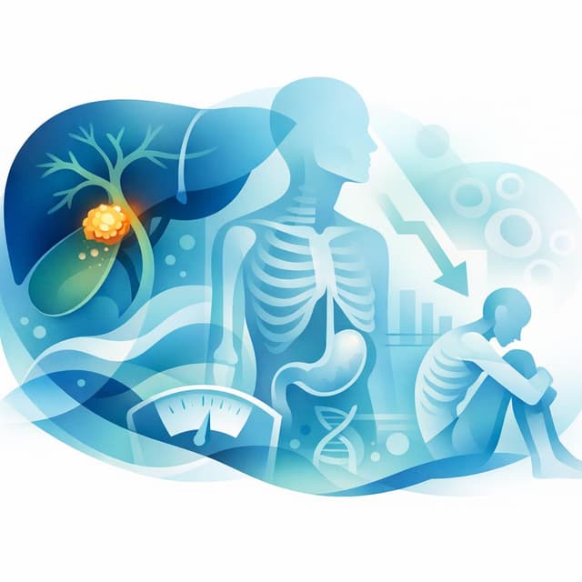
Nutrition and Weight-Loss Management
Cancer-related weight loss (malnutrition and cachexia) worsens outcomes; early intervention helps. Every evaluation should include a brief nutritional screen and plan. [14]
- Assess:
- Intervene:
- Dietitian referral, individualized counseling, and oral supplements to meet higher calorie and protein needs. [17]
- Consider appetite stimulants (e.g., megestrol acetate) for persistent anorexia after dietary measures. [15]
- Keep physically active as able to preserve muscle mass; consider multimodal care if cachexia suspected. [14]
For significant weight loss (e.g., ≥10%), stronger nutritional interventions and closer follow-up are generally recommended. [16]
Patient-Friendly Checklist
Use or adapt the following items for personal tracking and clinic visits. Bringing a completed checklist to appointments can speed up diagnostic steps. [1] [2]
-
Symptoms (check all that apply and note dates):
-
Weight and diet:
-
Medical history and risks:
-
Medications and lifestyle:
-
Evaluation steps discussed or completed:
Practical Testing Sequence at a Glance
- Start with labs (LFTs, CBC) and abdominal ultrasound for suspected biliary obstruction with weight loss. [11] [2]
- Proceed to contrast-enhanced CT or MRI/MRCP for better visualization and staging. [2] [6]
- Use EUS-FNA and/or ERCP for tissue diagnosis and biliary drainage when imaging suggests a malignant stricture or mass. [2]
- Consider PTC when ERCP is not feasible or anatomy precludes access. [10]
This sequence balances diagnostic accuracy with safety and provides a path to symptom relief (e.g., stenting) when obstruction is present. [2] [10]
Comparison Table: Symptoms and Diagnostic Steps
| Category | What to track or do | Why it matters |
|---|---|---|
| Jaundice, dark urine, pale stools, itching | Daily presence, onset, progression [1] [3] | Signals bile duct blockage that needs urgent evaluation [1] |
| RUQ pain, nausea/vomiting, fever/chills | Pain location/radiation, infection signs [2] [5] | Helps differentiate obstruction vs. infection and guides urgency [5] |
| Weight and diet | Weight trend, appetite, intake [7] [15] | Quantifies unintentional loss; triggers nutrition support [16] |
| Lab tests | Bilirubin, ALP, AST/ALT, CBC, CA 19-9, CEA [11] [2] | Detects cholestasis and supports suspicion of malignancy [6] |
| Imaging | Ultrasound → CT/MRI/MRCP [2] [6] | Maps biliary tree and characterizes strictures/masses [2] |
| Tissue diagnosis | EUS-FNA, ERCP brush/biopsy, PTC [2] [10] | Confirms malignancy and enables drainage if needed [2] |
When to Seek Care Urgently
- Rapidly worsening jaundice, fever/chills, or severe right-upper-quadrant pain can indicate cholangitis or acute obstruction. These symptoms warrant same-day medical attention. [5]
Bottom Line
Bile duct cancer often presents late because early signs are subtle; however, a focused checklist that captures jaundice, urine/stool color changes, itching, RUQ pain, and unintentional weight loss, paired with a stepwise workup (LFTs/CBC → ultrasound → CT/MRI/MRCP → EUS/ERCP with tissue sampling and possible drainage), can streamline evaluation and care. Integrating early nutritional assessment and support helps protect strength and treatment tolerance. [1] [11] [2] [6] [16] [14]
Related Questions
Related Articles
Sources
- 1.^abcdefghijklmnopqrBile Duct Cancer(medlineplus.gov)
- 2.^abcdefghijklmnopqrstuvwxyzDiagnosis of Cholangiocarcinoma.(pubmed.ncbi.nlm.nih.gov)
- 3.^abcdefghijklmCholangiocarcinoma: MedlinePlus Medical Encyclopedia(medlineplus.gov)
- 4.^abcdeBile Duct Cancer(nyulangone.org)
- 5.^abcdefghijBile duct stricture: MedlinePlus Medical Encyclopedia(medlineplus.gov)
- 6.^abcdefghijDiagnosis of cholangiocarcinoma.(pubmed.ncbi.nlm.nih.gov)
- 7.^abcWeight loss - unintentional: MedlinePlus Medical Encyclopedia(medlineplus.gov)
- 8.^abcdeWeight loss - unintentional: MedlinePlus Medical Encyclopedia(medlineplus.gov)
- 9.^abWeight loss - unintentional: MedlinePlus Medical Encyclopedia(medlineplus.gov)
- 10.^abcdef[Cholangiocarcinoma].(pubmed.ncbi.nlm.nih.gov)
- 11.^abcdefCholangiocarcinoma: MedlinePlus Medical Encyclopedia(medlineplus.gov)
- 12.^ab[Weight loss as a presenting clinical feature of malignancy].(pubmed.ncbi.nlm.nih.gov)
- 13.^↑Diagnosing Small Intestine Cancer(nyulangone.org)
- 14.^abcEarly recognition of malnutrition and cachexia in the cancer patient: a position paper of a European School of Oncology Task Force.(pubmed.ncbi.nlm.nih.gov)
- 15.^abcdAssessment and maintenance of nutrition in older cancer patients.(pubmed.ncbi.nlm.nih.gov)
- 16.^abcd[Good clinical practice in nutritional management in cancer patients: malnutrition and nutritional assessment].(pubmed.ncbi.nlm.nih.gov)
- 17.^↑The starving patient: supportive care for people with cancer.(pubmed.ncbi.nlm.nih.gov)
Important Notice: This information is provided for educational purposes only and is not intended to replace professional medical advice, diagnosis, or treatment. Always consult with a qualified healthcare provider before making any medical decisions.